
Autonomic Function & Exercise Intolerance
When the Patient Says, “I Can't Exercise Like I Used To,” Look Beyond Cardiac Output Alone.
Exercise intolerance is common across cardiovascular disease, but the causes are often multifactorial.
Reduced exercise capacity may involve cardiac reserve, pulmonary function, vascular responses, skeletal muscle function, conditioning, metabolic factors, medications, autonomic regulation, or combinations of these factors.
That makes exercise intolerance particularly important when the patient's symptoms appear greater than expected from the initial cardiovascular findings.
Physio PS is FDA 510(k) cleared and designed for use by physicians and healthcare providers.
The Autonomic Nervous System Is Part of the Exercise Response
Exercise requires rapid cardiovascular adaptation.
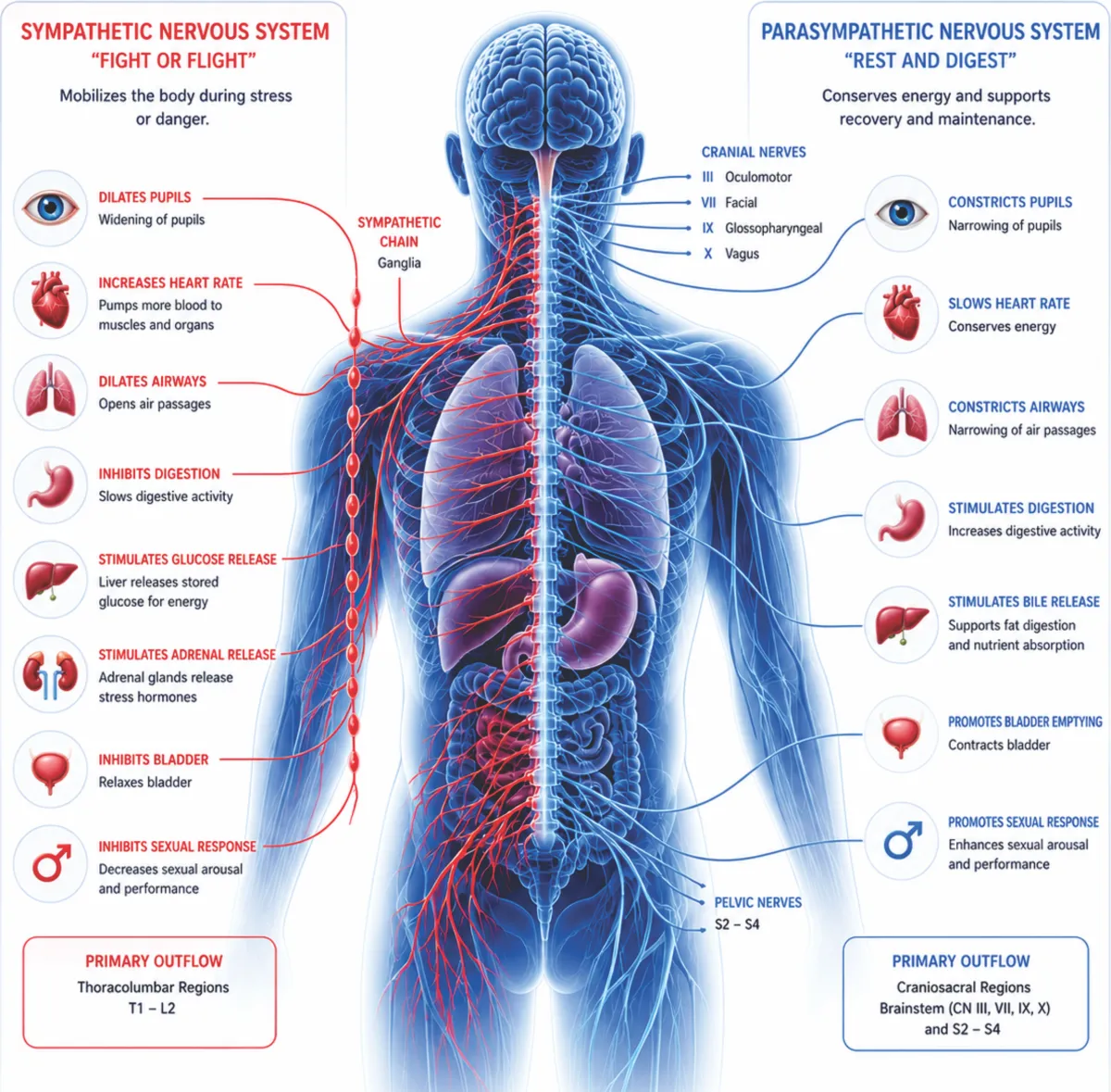
The autonomic nervous system contributes to:
Heart-rate acceleration
Vascular regulation
Redistribution of blood flow
Cardiovascular response to increased metabolic demand
Recovery following exertion
During exercise, parasympathetic activity normally withdraws while sympathetic activity increases. During recovery, parasympathetic activity increases again as cardiovascular demand decreases.
Abnormal autonomic responses have been associated with exercise intolerance in a number of clinical settings.
When Exercise Intolerance Raises Additional Questions
A cardiologist may encounter patients who report:
Reduced exercise capacity
Disproportionate fatigue with activity
Excessive heart-rate response
Poor heart-rate recovery
Lightheadedness during or after activity
Exercise-associated palpitations
Post-exertional worsening of symptoms
Exercise intolerance despite relatively reassuring conventional cardiac findings
These symptoms have many potential causes and require appropriate clinical evaluation.
Conventional Exercise Evaluation Still Matters
Depending on the patient, evaluation may include:
History and physical examination
ECG
Echocardiography
Exercise stress testing
Cardiopulmonary exercise testing
Rhythm monitoring
Laboratory evaluation
Pulmonary evaluation
Assessment of conditioning and other contributing factors
Autonomic assessment should complement—not replace—these established approaches when they are clinically indicated.

What Physio PS Adds
Physio PS provides quantitative information about parasympathetic and sympathetic activity.
The system's approximately 15-minute, non-invasive monitoring uses physiologic data including ECG, blood pressure and heart rate.
For selected patients with unexplained or disproportionate exercise intolerance, this may provide additional information about autonomic regulation that can be considered alongside the patient's broader cardiovascular assessment.
Why This Matters
Two patients can report the same symptom—“I can't exercise”—for very different physiologic reasons.
One may have reduced cardiac reserve.
Another may have pulmonary limitation.
Another may have deconditioning.
Another may have an abnormal autonomic response.
And some patients may have more than one contributing factor.
The Goal Is Not to Assign Every Exercise Symptom to the Autonomic Nervous System.
The goal is to determine whether autonomic physiology may be one part of the picture.
Establishing a Baseline
For selected patients, quantitative autonomic measurements may also provide a baseline that can be compared with subsequent assessments when clinically appropriate.
This can be useful when the clinician is monitoring physiologic changes over time as part of an overall treatment or rehabilitation strategy.
When Exercise Capacity Doesn't Match the Rest of the Clinical Picture, Consider the Regulatory System Behind the Response.
© Copyright 2026 Physio PS, Inc.-All rights reserved.